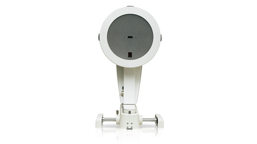
Overview
Within a mere second the Pentacam supplies you with precise diagnostic data on the entire anterior eye segment. The degree of cataract is made visible by the light scattering properties of the eye lens. Measurement of the anterior and posterior corneal surfaces supplies the true total refractive power of the cornea. The data on the posterior surface gives you just the assistance you need for early keratoconus detection.
The rotating scan supplies more data points in the centre of the cornea. A supplementary pupil camera corrects for eye movements during the examination. Unlike conventional topography systems the Pentacam measures true elevation data rather than just curvature values
Even the basic software offers a vast range of functions:
- Qualitative assessment of the cornea
- Topography and elevation data of the anterior and posterior corneal surfaces.
- Overall pachymetry, relative and absolute
- Glaucoma screening:
- Pachymetry-based IOP correction
- Chamber angle and volume
- Elevation data
- Topography assisted keratoconus detection and classification
- Comparative displays for follow-up
Need more functions? – No problem!
The Pentacam Basic can up front or later be upgraded to include the software packages for refractive surgery and/or cataract surgery and other optional software modules to suit your needs.
We’ll make your entry into the Pentacam world as easy as possible. With our new Pentacam Basic we have put together for you a configuration which offers all the basic functions you need for perfect diagnostics
With our modular software concept you can add more functional packages or analysis modules at any time.
This way your new Pentacam will grow with your demands
Scheimpflug image
Topography maps of the anterior and posterior corneal surface
Applications:
- Keratoconus detection
- Pre-surgical planning of refractive corneal surgery
- Follow-up after corneal surgery
- Calculation of IOL refractive power
- Planning of astigmatism reducing incisions (LRI)
- Follow-up after refractive surgery (pre-post LASIK)